


This is Issue 12 of CLI’s On Science Series.
Introduction
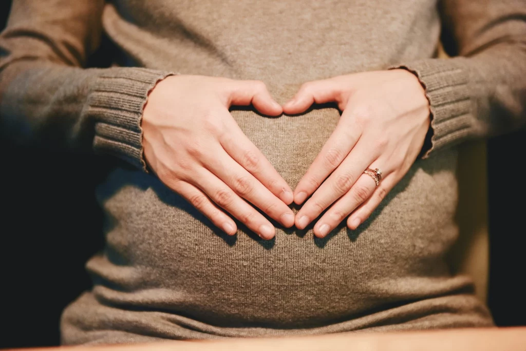
A unique human being forms at fertilization[1] when a man’s sperm fuses with a woman’s egg creating a zygote, a single-celled human embryo, that will become a 30-trillion cell adult.[2] From the moment of conception, a new human being contains a complete and unique set of genetic information that determines his or her physical traits, form, and range of abilities. There is enough genetic variation that no two humans have been, or ever will be, genetically identical.[3]
The genome of each unique human being is a blueprint or instruction book, ensuring that during development the heart beats at the right time, that eyes form on the front of the face, that bones grow inside the body, and that ears connect to the brain so that he can perceive his mother’s voice and the world around him. Every system in the body forms and functions according to a self-directed plan starting at fertilization and lasting for a lifetime.
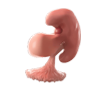
Ovulation and fertilization occur about two weeks after the woman’s last menstrual period (LMP). By three to four weeks, the developing embryo will have already traveled to the uterus and embedded into the uterine wall of his mother in a multi-day process called implantation. Almost immediately after implantation, the hormone called human chorionic gonadotropin (hCG) can be detected with over-the-counter urine pregnancy tests. And by six weeks, cardiac activity can be detected in the embryo by transvaginal ultrasound.
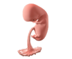
A new human being is called an embryo for the first 10 weeks of pregnancy and a fetus from 11 weeks until birth. The age of an unborn child can be calculated two different ways. Doctors usually date a pregnancy from the start of the mother’s last menstrual period (LMP), called the gestational age. But pregnancy biologically starts at fertilization. So, if a doctor tells a woman that she is 12 weeks pregnant, then that means that the unborn child has been developing inside her mother’s womb for about 10 weeks since sperm-egg fusion (fertilization).
Below we describe 12 amazing facts about the developing human being at 12 weeks of gestation. Enjoy the journey!
12 Amazing Facts about the Developing Human Being at 12 Weeks of Gestation
1) All of the major organs have formed.
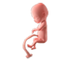
Almost every organ and tissue form by 12 weeks. The remainder of the pregnancy is spent growing these organs larger and more mature to prepare for life outside the womb.[4] By 10 weeks’ gestation, she will already have over 90% of the more than 4,500 named structures in the adult body including arms, legs, fingers, toes, a face, and eyelids.[5] The digestive system, skin, and muscles have all started to function. The fetus may still be small at 12 weeks (about 2.5 inches in length weighing less than an ounce), but she is already an incredibly complex and multi-faceted human being!
2) The four-chambered heart pumps over six quarts of blood per day.
The four chambers of the heart form by the eighth gestational week.[6] The heart reaches its final shape by 10 weeks. The heart is a vital source for circulation of nutrients and oxygen-carrying blood.[7] The heart and circulatory system of a 12-week-old fetus pumps an average of six quarts of blood per day, which is equivalent to one-and-a-half gallons of milk. For comparison, an adult heart pumps 6,000 quarts of blood each day.[8]
3) The heart has already beat over 10 million times.
About 22 days after fertilization (sixth week of gestation), the heart starts to beat rhythmically.[9] The heart beats around 110 beats per minute during the sixth week, increasing to approximately 160 beats per minute by eight weeks, and peaking during the ninth week to twice the heart rate of the mother’s at over 170 beats per minute.[10] The average heart rate at 12 weeks is 167 beats per minute. By then, the heart has already beat over 10 million times.[11]
4) Each finger moves separately.
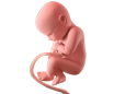
Starting at 10-and-a-half weeks’ gestation, when something touches the fetus’s hand, he starts to close his fingers.[12] By 10 weeks, he will bring his hands together and touch his face. Typically, the fetus moves all of his fingers together, except the thumb. Over the next few weeks, he starts to bend his fingers more deeply and move his thumb, as if he were grasping an object. By 12 weeks’ gestation, the fetus moves each finger separately and spontaneously explores his environment with his fingers.[13]
5) The fetus sucks his thumb, with a preference for his left or right hand.
As early as 10 weeks’ gestation, it is possible to determine whether the unborn child is left-handed or right-handed by studying ultrasounds. About 85% of fetuses prefer moving their right hand over their left hand, and about 85% of adults prefer their right hand, too.[14] When examining the same children over time, almost every fetus that preferred sucking his or her right thumb remained right-handed, but only a few of the fetuses that sucked their left thumbs in their mother’s womb changed preference and were right-handed by the age of 10.[15]
6) Unique fingerprints are forming.
Every human being has unique fingerprints, which start forming during the 12th week of gestation. As a subset of cells in the skin start growing faster than other cells, these extra cells cause the skin to buckle and fold into ridges.[16] As the fingers grow, new ridges and branches form in the fingerprints. Later in development, a layer of keratin coats the surface of the skin and smaller, secondary ridges form out of uneven keratin growth. The fetal environment in the womb can influence fingerprint pattern including the fetus’s size, location, and movement patterns.[17] The individual fingerprint patterns are established by 19 weeks and remain consistent as the child grows.[18]
7) The fetus has a face.
By the 12th week of gestation, the fetus has a recognizable face with prominent facial features including a forehead, nose, cheeks, jaw, tongue, upper and lower lips, eyes, and ears.[19]
8) The fetus exhibits complex behaviors.
By 12 weeks’ gestation, the fetus shows complex behaviors such as yawning, stretching, swallowing, scrunching hands into little fists, touching her face, and scratching her head.[20] The fetus can now bend her elbows and bring her hands together, squint, grasp and point her toes.[21] She even makes intermittent breathing motions.[22]
9) Teeth are developing.
Teeth start to develop as buds in the ninth week of gestation. These first teeth to form become the “baby teeth” that start to poke out of the gum starting roughly six months after birth.[23] Most children have their full set of 20 baby teeth by age 2. Around 11 weeks’ gestation, the buds of the permanent teeth start to develop underneath the baby teeth.[24] This second set of “adult teeth” will slowly develop into the permanent teeth of an adult that replace the baby teeth throughout childhood.
10) The body responds to touch and may experience pain.
Touch receptors can be seen around mouth and hands at seven-and-one-half weeks’ gestation,[25] and the embryo starts to reflexively move away at eight weeks.[26] Nerve synapses for spinal reflex are completely in place by 10 weeks.[27] For example, when something touches the bottom of the foot, a 12-week-old fetus will curl his or her toes,[28] just like the adult reflex.
Pain receptors (nociceptors) begin forming at seven weeks’ gestational age,[29] with the nerves linking pain receptors to the pain-sensing part of the brain, the thalamus, forming at 12 weeks.[30] A comprehensive review of the scientific literature including neural development, psychology of pain sensation, and moral implications of fetal pain, concludes that unborn babies may experience pain as early as 12 weeks.[31]
11) The brain is forming at a rapid rate.
The brain and spinal cord start forming in the fifth week of gestation.[32] As the pregnancy progresses, the brain grows and folds to create distinct brain regions. To support this growth, neurons grow fast — the brain creates over 250,000 neurons per minute. Neural circuits, or connections between neurons that communicate information throughout the brain, form at a rapid rate in the fetal period. Neurons start producing chemical messengers that allow them to communicate right away, and spontaneous electrical activity has been recorded from connected neurons as early as the eighth week of gestation.[33] More and more brain areas become fully functional as the fetus matures and is born. Nevertheless, the brain is not fully mature until about 25 years of age.
12) Brain connections formed at 12 weeks’ gestation survive into adulthood.
Sensory information from the eyes, ears, mouth, and body travels via nerves to subcortical areas and a brain structure called the thalamus, the gateway to the cortex. The thalamus acts like a relay center, condensing some information, or reducing a signal, if for instance the person is asleep, before passing it on to the cerebral cortex. The cerebral cortex helps process emotions, decision-making, working memory and attention. Many abilities that make human beings unique and distinct from other animals are governed by the cerebral cortex. Before the cerebral cortex forms, a thick plate of neurons forms, called the subplate.[34] Neurons destined for the cerebral cortex first migrate into the subplate while they wait for the supporting cells in the cortex to mature. Then they migrate into their final positions. Eventually, the subplate fades away and becomes white matter.[35]
Scientists have found connections between the thalamus and the subplate as early as 12 weeks’ gestational age.[36] Recent work has shown that connections between the auditory nerves, auditory region in the thalamus, and neurons in the subplate survive into adulthood.[37] Auditory information is organized by pitch in the ear, thalamus, and cortex. That organization pattern was also seen in the subplate, suggesting that preliminary high-level processing may happen long before the cortex is fully formed.[38]
Conclusion
If one follows the science, it’s clear that preborn children at 12 weeks’ gestation are already amazingly complex human beings. All of the major internal organs are formed and are already functioning, including the heart. They start forming brain connections that will last into adulthood, are developing unique fingerprints, and already show a preference for their right or left hand. To minimize or ignore the humanity of the 12-week-gestation fetus is to deny the clear and compelling evidence of science.
[1] Ronan O’Rahilly and Fabiola Müller, Developmental Stages in Human Embryos: Including a Revision of Streeter’s “Horizons” and a Survey of the Carnegie Collection (Washington D.C.: Carnegie Institution of Washington, 637, 1987); and Sadler, Thomas W., Medical Embryology, 14th edition, 2019; and Dr. Maureen Condic, Charlotte Lozier Institute, On Point “A Scientific View of When Life Begins”, available at: https://lozierinstitute.org/a-scientific-view-of-when-life-begins/, June 2014.
[2]AC Guyton and JE Hall, Textbook of Medical Physiology., 10th ed. (Philadelphia: W.B. Saunders, 2000).
[3] National Institutes of Health (US) and Biological Sciences Curriculum Study, Understanding Human Genetic Variation, NIH Curriculum Supplement Series [Internet] (National Institutes of Health (US), 2007), https://www.ncbi.nlm.nih.gov/books/NBK20363/.
[4] Sadler, Thomas W., Medical Embryology, 14th edition, 2019.
[5] The Endowment for Human Development. Carnegie Stage 23 Introduction. Available at: https://www.ehd.org/virtual-human-embryo/intro.php?stage=23
[6] Tan C, M, J, Lewandowski A, J: The Transitional Heart: From Early Embryonic and Fetal Development to Neonatal Life. Fetal Diagn Ther 2020;47:373-386.
[7] Hill, M.A. (2023, January 28),Cardiovascular System Development, Available at: https://embryology.med.unsw.edu.au/embryology/index.php/Cardiovascular_System_Development
[8] F. S. Molina et al., “Heart Stroke Volume and Cardiac Output by Four-Dimensional Ultrasound in Normal Fetuses,” Ultrasound in Obstetrics & Gynecology: The Official Journal of the International Society of Ultrasound in Obstetrics and Gynecology 32, no. 2 (August 2008): 181–87, https://doi.org/10.1002/uog.5374.
[9] Sadler, Thomas W., Medical Embryology, 14th edition, 2019. Page 181; and Hill, M.A. (2023, January 28), Cardiovascular System Development, Available at: https://embryology.med.unsw.edu.au/embryology/index.php/Cardiovascular_System_Development
[10] Murugan VA, Murphy BO, Dupuis C, Goldstein A, Kim YH. Role of ultrasound in the evaluation of first-trimester pregnancies in the acute setting. Ultrasonography. 2020 Apr;39(2):178-189. doi: 10.14366/usg.19043. Epub 2019 Oct 16. PMID: 32036643; PMCID: PMC7065984; and Hornberger LK, Sahn DJ. Rhythm abnormalities of the fetus. Heart. 2007 Oct;93(10):1294-300. doi: 10.1136/hrt.2005.069369. PMID: 17890709; PMCID: PMC2000955.
[11] EHD: Appendix, “Appendix | Prenatal Overview,” accessed March 15, 2023. https://www.ehd.org/dev_article_appendix.php#fb16.
[12] Humphrey, T., “Some Correlations between the Appearance of Human Fetal Reflexes and the Development of the Nervous System,” in Progress in Brain Research, ed. Dominick P. Purpura and J. P. Schadé, vol. 4, Growth and Maturation of the Brain (Elsevier,1964), 93–135, https://doi.org/10.1016/S0079-6123(08)61273-X.
[13] Ibid.
[14] Peter G Hepper, Glenda R McCartney, and E. Alyson Shannon, “Lateralised Behaviour in First Trimester Human Foetuses,” Neuropsychologia 36, no. 6 (June 1, 1998): 531–34,https://doi.org/10.1016/S0028-3932(97)00156-5.
[15] Peter G. Hepper, Deborah L. Wells, and Catherine Lynch, “Prenatal Thumb Sucking Is Related to Postnatal Handedness,” Neuropsychologia 43, no. 3 (January 1, 2005): 313–15,https://doi.org/10.1016/j.neuropsychologia.2004.08.009.
[16] Babler, W. J. “Embryologic Development of Epidermal Ridges and Their Configurations.” Birth Defects Original Article Series 27, no. 2 (1991): 95–112. “Embryologic Development of Epidermal Ridges and Their Configurations.”
[17] Singh, RK, M Sharma, A Tarannum, W Pet-Paul, and L Bernard. “A Case of Three Deltas in a Fingerprint.” J Forensic Res 9, no. 413 (2018): 2. https://doi.org/10.4172/2157-7145.1000416.
[18] Babler, “Embryologic Development of Epidermal Ridges and Their Configurations.”
[19] Sadler, Thomas W., Medical Embryology, 14th edition, 2019.
[20] Liley, A. W. “The Foetus as a Personality.” The Australian and New Zealand Journal of Psychiatry 6, no. 2 (June 1972): 99–105. https://doi.org/10.3109/00048677209159688.
[21] de Vries, J. I. P. de, G. H. A. Visser, and H. F. R. Prechtl. “The Emergence of Fetal Behaviour. I. Qualitative Aspects.” Early Human Development 7, no. 4 (December 30, 1982): 301–22. https://doi.org/10.1016/0378-3782(82)90033-0.
[22] Ibid.
[23] Sadler, Thomas W., Medical Embryology, 14th edition, 2019.
[24] Ibid.
[25] Humphrey, T., “Some Correlations between the Appearance of Human FetalReflexes and the Development of the Nervous System,” in Progress in Brain Research, ed.Dominick P. Purpura and J. P. Schadé, vol. 4, Growth and Maturation of the Brain (Elsevier,1964), 93–135, https://doi.org/10.1016/S0079-6123(08)61273-X.
[26] Hooker, D. The Prenatal Origin of Behavior. Lawrence, KS: University of Kansas Press, 1952.
[27]Okado N et al., Synaptogenesis in the cervical cord of the human embryo: Sequence of synapse formation in a spinal reflex pathway, J. Comparative Neurol. 184, 491, 1979; Okado N, Onset of synapse formation in the human spinal cord, J. Comparative Neurol. 201, 211, 1981.
[28] Humphrey, T., “Some Correlations between the Appearance of Human Fetal Reflexes and the Development of the Nervous System.”
[29] Myers LB et al., Fetal endoscopic surgery: indications and anaesthetic management, Best Pract Res Clin Anaesthesiol 18, 231, 2004, doi: 10.1016/j.bpa.2004.01.001; Anand KJS, Hickey PR. Pain and its effects in the human neonate and fetus. New England Journal of Medicine. 317, 1321-1329, 1987, doi: 10.1056/NEJM198711193172105
[30] Derbyshire SWG and Bockmann JC, Reconsidering Fetal Pain, Journal of Medical Ethics 46, 3-6, 2020, doi: 10.1136/medethics-2019-105701; Kostović I, Judaš M, The development of the subplate and thalamocortical connections in the human foetal brain, Acta Paediatr 99, 1119–1127, 2010, doi: 10.1111/j.1651-2227.2010.01811.x; Van de Velde M, De Buck F, Fetal and Maternal Analgesia/Anesthesia for Fetal Procedures, Fetal Diagnosis and Therapy 31, 201, 2012, doi: 10.1159/000338146
[31] Derbyshire and Bockmann, “Reconsidering Fetal Pain.”
[32] Sadler, Thomas W., Medical Embryology, 14th edition, 2019.
[33] Borkowski, Winslow J., and Richard L. Bernstine. “Electroencephalography of the Fetus.” Neurology 5, no. 5 (May 1, 1955): 362. https://doi.org/10.1212/WNL.5.5.362.
[34] Anna Hoerder-Suabedissen and Zoltán Molnár, “Development, Evolution and Pathology of Neocortical Subplate Neurons,” Nature Reviews Neuroscience 16, no. 3 (March 2015):133–46, https://doi.org/10.1038/nrn3915.
[35] Stuart and Bockmann, “Reconsidering Fetal Pain.”
[36] Ivica Kostović and Nataša Jovanov-Milošević, “The Development of Cerebral Connections during the First 20–45 Weeks’ Gestation,” Seminars in Fetal and Neonatal Medicine, Assessing Brain Function in the Perinatal Period, 11, no. 6 (December 1, 2006):415–22, https://doi.org/10.1016/j.siny.2006.07.001.
[37] Jessica M. Wess et al., “Subplate Neurons Are the First Cortical Neurons to Respond to Sensory Stimuli,” Proceedings of the National Academy of Sciences 114, no. 47 (November21, 2017): 12602, https://doi.org/10.1073/pnas.1710793114.
[38] Derbyshire and Bockmann, “Reconsidering Fetal Pain.”